Flow cytometry is a powerful analyzing technique based on the production of an electrical signal proportional to the physical and chemical properties of a cell or another particle in a fluid medium.
Immunology, oncology, and clinical diagnostics are some sectors where this technology has a major utility. Thus, the realization of a powerful tool for rapid quantitative data on cells has introduced flow cytometry into modern biological practice and changed the approaches to investigating numerous problems in both basic and clinical research.
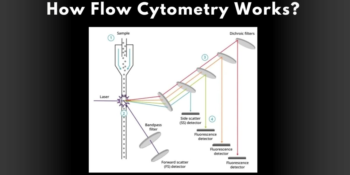
Flow cytometry in its simplest form involves the irradiation of cells in suspension with a laser beam. Illuminating cells with the aid of a beam allows the light to scatter and a cell will fluoresce if it has been tagged with fluorescent dyes. This scattered light and fluorescence is what a flow cytometer can then use to measure a variety of characteristics regarding each cell including the size, the granularity, and the nature of proteins within it. This process enables finding the characteristics of thousands of cells in one second so that it may be considered an efficient procedure in characterizing cells.
Key Components of Flow Cytometry
Several key components contribute to the functionality of a flow cytometer:
- Fluidics System: This system is utilized to convey cells suspended in a fluid flow towards the laser beam and is mandatory for the correct operation of the whole apparatus. It makes certain that a cell file is well aligned hence may easily be interacted with by the laser.
- Laser: The centerpiece of the flow cytometer is the laser. It supplies the kind of light that will precipitate the fluorescence of the labels attached to the cells. This information shows that the available laser may excite various markers in the fluorescent assays so that multiple assays may be run in parallel.
- Detectors: The light emitted by the cells that pass through the laser beam is finally collected by a chain of photodetectors. These detectors quantify the density of scattered light and fluorescence produced by each cell. The collected data are then translated into computer-usable form for analysis.
- Computer Software: The detectors assemble different data analyzed using sophisticated software. Such tools may reveal the distribution and other parameters of the population of cells of interest or their concrete features.
The Flow Cytometry Process
The flow cytometry process may be broken down into several steps:
- Sample Preparation: The first flow cytometry procedure is sample preparation. Cells have to be proved in an ideal buffer and often in order need to be labelled with antibodies conjugated with fluorides that bind separate markers. Sample preparation is very important as it may impact the overall result.
- Loading the Sample: After preparation, the sample is then loaded into the flow cytometer. Flow cytometry is completed with a collection of four protocols. The fluidics system aspirates the sample into the instrument and positions the cells in a stream since most particle analysis requires the identification of particles in a stream.
- Excitation and Detection: Cellular response to the laser beam is achieved by passing the cells through it and capturing the light emitted by passed cells. The system includes forward scatter, which corresponds to cell size, side scatter, which corresponds to cell granularity, and fluorescence from tagged markers.
- Data Analysis: This detection is followed by the signal transmitting to the computer software where an assessment of the signals takes place. Using the software, various types of plots such as histograms, dot plots, and other formats may be obtained for ease of understanding of the outcomes.
- Interpreting Results: The analyzed data is then further analyzed to come up with significant conclusions on the cell populations. They may DISSECT this analysis for specific details regarding the health, functioning, and identity of specific cells which would be Central to various studies and therapy plans.
Applications of Flow Cytometry
Flow cytometry plays a wide role in immunology, oncology, hematology, microbiology, genetics, evolution, astrophysics, and other many disciplines.:
- Immunology: Flow cytometry was used to investigate immune cell population, their activation, and response to treatments by researchers. That is why it is critical to institute diseases and work on the corresponding treatment methods.
- Cancer Research: In oncology, flow cytometry is used to identify the characteristics of all tumors, measure MRD, and determine the efficacy of therapeutic approaches. It reveals some information regarding the variation of tumors.
- Clinical Diagnostics: Flow cytometry is a routine diagnostic method in most clinical laboratories and is applied for the diagnosis of blood diseases including leukemia and lymphoma. It helps in the management of the severity of an illness and how it should be managed.
- Stem Cell Research: The use of flow cytometry instrumentation in the isolation and Characterization of stem cells is useful in research for regenerative medicine and tissue engineering.
The Advantages and Disadvantages of Flow Cytometry
Flow cytometry offers several advantages:
- Speed: Flow cytometry has the advantage of generating results at great speed with thousands of cells analyzed per second.
- Multiplexing: Several fluorescent markers may work at once allowing sampling of more than one parameter in a given sample.
- Quantitative Data: This technology gives clues that give quantitative data as the samples are compared.
However, flow cytometry also has limitations:
- Sample Preparation: Sample preparation is important, and any mistake that one makes to do so will only give him or her the wrong result.
- Cost: Some flow cytometers are costly, and performing the assays demands costly reagents and pulling-through training.
- Complexity: Flow cytometry data analysis may be challenging with many possibilities for misinterpretation which requires understanding to avoid.
Future Directions in Flow Cytometry
Technological development has not left flow cytometry out, and there are constant improvements. Technological advancements including spectral flow cytometry are preferable because they enable a simultaneous identification of even more fluorescent labels leading to increased possibilities of analysis. In addition, combining with other methods, for example, mass cytometry or single-cell RNA sequencing, is expanding new opportunities.
Flow cytometry has become an essential technique in present biological and medical studies. This technique has accelerated our ability to study and quantify the functions and dysfunctions of single-cell organisms and complexes.
As there is a constant development of new technology and an increased knowledge of the human cell, flow cytometry is likely to continue to be the leading biological tool for examining new discoveries and diagnostics.
To find out more about what flow cytometry is, as well as its uses, flow cytometry animation and various other articles, please visit Abcam.
Disclaimer: This article is intended for informational purposes only and does not constitute professional or medical advice. While efforts have been made to ensure the accuracy of the information, readers are encouraged to consult relevant professionals for specific advice or guidance. Abcam is not liable for any errors or omissions in this content or for any actions taken based on the information provided.
Published by Tom W.


